|
|
Blount Disease
Tibia Vara
General Considerations
- Growth disturbance in the proximal, medial, epiphyseal plate, epiphysis, and metaphysis of the tibia produces medial angulation (varus deformity) at the knee and internal rotation of the tibia
- Pathogenesis is uncertain
- The early onset or infantile type occurs in children under 3 years of age (more common)
- The later-onset occurs in a juvenile (4-10) and adolescent form (11 and older)
- Infantile form is more common in females, African-Americans and the overweight
- Adolescent form also is more common in overweight obese
Clinical Findings
- Infantile form is usually free of pain whereas adolescent form is accompanied by pain
- Infantile form is bilateral in 80% of cases and associated with greater deformity
- Adolescent form is unilateral in 80% and associated with less deformity
Imaging Findings
- The study of choice is a standing AP view of the knees and a lateral view
- Infantile form is bilateral in 80% of cases and associated with associated with a prominent metaphyseal beak
- Internal tibial torsion
- Leg-length discrepancy (shorter on affected side)
- Fragmentation of the medial epiphysis which is wedge-shaped
- Varus deformity is measure by the tibiofemoral angle
- Severity of changes are assessed by using the metaphyseal-diaphyseal angle of the tibia
- Angle is formed by lines between metaphyseal beaks and perpendicular to the longitudinal axis of the tibia; >11 degrees is abnormal
Differential Diagnosis
- Marked physiologic bowlegs
- Common in toddlers who walk at an early age and are overweight; usually resolves spontaneously by age 2 years
Treatment
- Orthotic management includes long-leg, locked-knee braces
- Indications for operative treatment include increasing severity of symptoms or progression of deformity
Prognosis
- Adolescent Blount disease does not appear to be as progressive infantile form

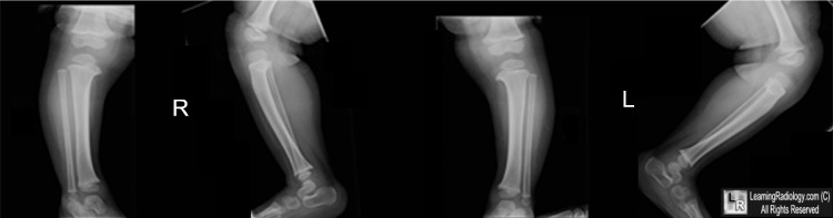
Bilateral Blount Disease. Frontal and lateral radiographs of both the right and left lower legs show beaking of the medial tibial plateau (white arrows), downward angulation of both proximal tibias. The tibiofemoral angle (blue lines)shows a varus deformity and the metaphyseal-diaphyseal angle is greater then 11 degrees (yellow lines).
For this same photo without arrows, click here
For more information, click on the link if you see this icon 
|
|
|
